New Year’s Holiday Sale: ends december 31st
.png)
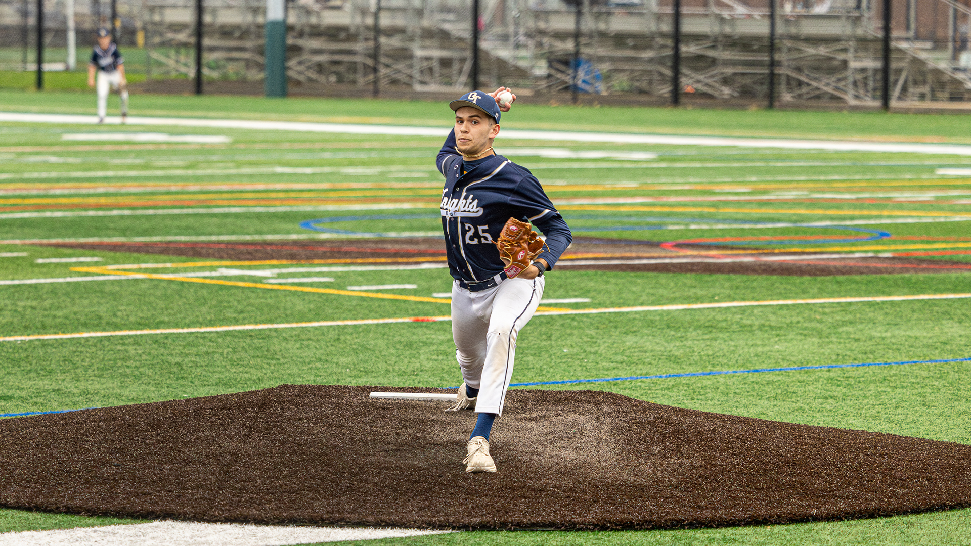
Every spring, a new crop of high school pitchers walks into training facilities across the country chasing the same target—velocity. It is measurable, visible, and glorified. But behind that radar-gun obsession sits an uncomfortable truth: elbow injuries in adolescent throwers are still climbing, and many of those athletes were completely pain-free just weeks before breaking down.
In 2025, Ryosuke Nishi and colleagues published a prospective study that may help explain why. Over six months, they tracked 128 high school baseball players, all symptom-free at baseline, using a combination of ultrasound imaging, joint mobility testing, and wearable technology to monitor throwing metrics. Their question was simple but rarely answered with this level of precision: what actually predicts medial elbow injury in adolescent pitchers?
What they discovered disrupts much of the conventional wisdom. The best predictor of who got hurt wasn’t velocity, torque, or arm slot. It was what the bone looked like. Subtle structural changes at the medial epicondyle—specifically irregularity and hypertrophy—carried odds ratios of 5.4 and 3.2 for future injury. And even more surprising, these irregularities were present in many “healthy” athletes before a single throw of the season.
Baseball culture has always prized quantifiable performance—velocity, spin rate, torque readings, workload counts. They create an illusion of precision, as if every ounce of stress can be monitored and managed in real time. Yet year after year, elbow injuries occur in athletes whose wearable data shows nothing unusual.
The deeper issue is biological. High school athletes occupy an unstable intersection of growth and load. Their skeletons are not yet fully mature, and the medial epicondyle—where the ulnar collateral ligament (UCL) anchors—is particularly vulnerable to traction stress. The growth plate in this region closes late, and before it does, repetitive valgus load creates microtrauma that gradually reshapes the bone.
Nishi’s study gives that concept objective weight. By imaging every athlete’s elbow before the season began, the authors could separate existing structural change from functional mechanics or acute overload. In doing so, they exposed a critical blind spot in how most programs evaluate readiness: physical screens focus on strength, range, and mechanics, but rarely on the integrity of the underlying tissue.
Even more striking was the sheer prevalence of prior injury. Nearly 70 percent of athletes reported a history of elbow pain despite being cleared for full participation. That statistic reframes the problem. Injury risk in young throwers is not a binary—healthy or hurt—but a continuum of accumulated adaptation where the tissue may look healed but remain fundamentally altered.
Across the six-month observation window, 23 of the 128 pitchers (18 percent) developed elbow pain lasting at least one week. When the data was modeled, three predictors emerged:
Notably absent from the list were all the mechanical parameters tracked through the Motus BASEBALL sleeve—elbow torque, arm slot, and arm speed. None showed a significant correlation with future pain.
That last point is critical. Wearable data has become a cornerstone of load management systems, but this study suggests it may only describe momentary stress, not tissue capacity. Torque does not predict breakdown if the structure beneath it has already been remodeled to tolerate—or fail under—certain thresholds.
The two anatomic findings tell complementary stories.
Both indicate that the medial elbow is already remodeling in response to prior load. These features do not guarantee pain, but they mark a joint that has been through significant mechanical history. In other words, the elbow “remembers.”
The third factor—greater shoulder external rotation—is a more nuanced signal. Increased ER has long been associated with velocity development, but in immature athletes it also amplifies valgus torque downstream. Each additional degree of layback increases the lateral distraction and medial compression on the elbow. For a 15- to 17-year-old whose epicondyle is still ossifying, that mechanical advantage can become structural overload.
The implication is not that throwing hard or having large ER range is inherently dangerous. Rather, it means that developmental context determines whether adaptation is functional or degenerative. A mature professional pitcher may use that same layback safely because his skeletal architecture has already stabilized.
At Velo University, we use findings like this to reframe what “readiness” actually means. Traditional screens prioritize motion and strength symmetry. But if an athlete presents with full range, strong cuff, and efficient mechanics yet has a history of medial pain, the next question is structural—what does the tissue look like?
While we may not have a diagnostic ultrasound in every training bay, the underlying logic is transferable. Three operational principles emerge from Nishi’s work:
These applications align with a broader philosophy: prevention is not about suppressing output but synchronizing it with what the athlete’s structure can currently tolerate.
The Nishi study dismantles the comforting assumption that performance metrics alone can forecast injury. For adolescent pitchers, the earliest warning signs may be invisible to the naked eye—a thickened growth plate, a subtly irregular bone surface, or a shoulder that rotates just a bit too freely for its own stability.
In practice, this means that prevention must evolve from a reactive to a predictive science. Rather than asking, how much stress can this arm handle today?, the better question is, what does this arm’s history tell us about its capacity to adapt tomorrow?
For coaches and clinicians, the take-home is both humbling and empowering. Wearables, motion capture, and high-speed video offer real-time feedback, but they must be interpreted through the lens of tissue health and developmental stage. Strength and mobility matter—but only if the structure beneath them is prepared to bear the load.
At Velo University, this shapes how we evaluate every athlete entering the building. We treat prior injury as a starting point, not a footnote. We measure not just how hard they can throw, but how efficiently they can recover. Because the true marker of longevity isn’t the peak torque number or radar reading—it’s how quietly the elbow behaves over time.
The study ends with a cautious note: identifying these structural risk factors is only the first step. The next challenge is figuring out whether early detection and targeted intervention can change the trajectory. That is where applied baseball science must go next—bridging diagnostic insight with developmental strategy.
Velocity will always be the headline metric. But if we want more pitchers to make it through adolescence healthy enough to keep chasing it, we have to start reading the quieter signals underneath.
Nishi, R., Ogawa, T., Obokata, Y., Furushima, K., & Kamatani, K. (2025). Anatomic, functional, and mechanical risk factors for elbow injury in the throwing athlete: A prospective cohort study of 128 high school baseball players. The American Journal of Sports Medicine, 53(4), 1462–1470. https://doi.org/10.1177/03635465251326579

.png)
.png)